
12. tbl. 101. árg. 2015
Decellularized fish skin: characteristics that support tissue repair
Introduction: Acellular fish skin of the Atlantic cod (Gadus morhua) is being used to treat chronic wounds. The prevalence of diabetes and the comorbidity of chronic wounds is increasing globally. The aim of the study was to assess the biocompatibility and biological characteristics of acellular fish skin, important for tissue repair.
Materials and methods: The structure of the acellular fish skin was examined with microscopy. Biocompatibility of the graft was conducted by a specialized certified laboratory. Protein extracts from the material were analyzed using gel electrophoresis. Cytokine levels were measured with an enzyme linked immunosorbent assay (ELISA). Angiogenic properties were assessed with a chick chorioallantoic membrane (chick CAM) assay.
Results: The structure of acellular fish skin is porous and the material is biocompatible. Electrophoresis revealed proteins around the size 115-130 kDa, indicative of collagens. The material did not have significant effect on IL-10, IL-12p40, IL-6 or TNF-α secretion from monocytes or macrophages. Acellular fish skin has significant effect on angiogenesis in the chick CAM assay.
Conclusion: The acellular fish skin is not toxic and is not likely to promote inflammatory responses. The graft contains collagen I, promotes angiogenesis and supports cellular ingrowth. Compared to similar products made from mammalian sources, acellular fish skin does not confer a disease risk and contains more bioactive compounds, due to less severe processing.
Table I. Overview of biocompatibility tests. All tests were conducted by Toxicon except the endotoxin test, that was conducted by Isotron Laboratories.
| Test description | Organism | Results | |
| MTT test: MTT (3-(4,5-imetylþiasol-2-yl)-2,5-difenyltetrasolium) with acellular fish skin was dissolved in medium (3 cm^2/mL) and placed in a cell culture for 24 hours. Degradation of MTT was assessed with spectrometry. | L929 fibroblasts from mouse(strain: C3H) | Acellular fish skin did not have effect on the survival of L929 fibroblasts: 79% of cells were alive after treatment with the fish skin compared to negative control. Tested according to ISO 10993-5. | |
| MEM test: MEM (Minimum Essential Medium) with fish skin (3 cm^2/mL) was added to a confluent cell culture for: 0, 24 or 48 hours. Shape of the cells was assessed for toxicity. | L929 fibroblasts from mouse(strain: C3H) | Acellular fish skin did not affect the shape of L929 fibroblasts. Tested according to ISO 10993-5. | |
| Systemic toxicity test: Fish skin (30 cm^2) was dissolved and administrated (2 mL/min, ~≤50 mL/kg). The mice were weighted before the received the fish skin and after: 24, 48 and 72 hours. | Albino mice, Switzerland | Acellular fish skin administrated to mice did not show signs of toxicity. All mice showed normal body weight gains and no signs of toxicity. Tested according to ISO 10993-5. | |
| Intramuscular grafting: Fish skin (1X10 mm strips)was placed in paravertebral muscle of mice for 1 week and assessed with H&E staining and microscopy. | Albino mice, Switzerland | Intramuscular grafting of acellular fish skin did not exhibit signs of toxicity and showed minor signs of inflammation. Tested according to ISO 10993-5. | |
| Pyrogenicity test: Dissolved fish skin (3 cm^2/mL) was administrated (~≤10 ml/kg) to rabbits. Body temperature was measured before and after and then every 30 minutes after injection. | White rabbits, New Zealand | Acellular fish skin did not cause fever in rabbits. Body temperature did not rise above 0,5°C in none of the rabbits tested. Tested according to ISO 10993-11. | |
| Endotoxin test: Fish skin (21cm^2) was placed in 200 mL of water for 15 minutes at 37°C. Amount of endotoxin (Eu/mL) was then measured in the water. | - | Amount of endotoxin was within given standards (20EU/test material). | |
| Irritation test: Fish skin (6,25 cm^2) was placed on a rabbit skin for 4 hours. Then the skin are was monitored: 1, 24, 48 and 72 hours after the removal of the fish skin. | White rabbits, New Zealand | Acellular fish did not cause inflammation on rabbit skin. Skin that was in direct touch with the fish skin did not show any signs of inflammation after 72 hours. Tested according to ISO 10993-10. | |
| Buehler sensitization test: Fish skin (6,25 cm^2) was placed on a skin for 6 hours. The skin area was monitored 24 and 48 hours after the removal of the fish skin. | Albino hamster | Acellular fish skin did not increase sensitivity of hamster skin according to given standards. Tested according to ISO 10993-10 | |
| Ames test: Bacterial strains unable to form histidine or tryptophan were placed in medium without these amino acids, with or without fish skin (3 cm^2/mL) and plated. Plates were incubated at 37±1°C and after 70 hours they were assessed for colonies. | Salmonella typhimurium and Escherichia coli | Acellular fish skin did not show increase rate of mutations compared to negative control (p ≤ 0,05). Tested according to ISO 10993-3 | |
| Chromosomal aberration assay. Fish skin was dissolved (3 cm^2/mL), placed in cell culture and removed after 3 hours. The cells were allowed to grow for 24 hours and then stained with Giems stain that binds chromosomes. | Chinese hamster ovary cells |
Acellular fish skin did not cause changes in chromosome shape compared to negative control. Tested according to ISO 10993-12 | |
| Micronucleus assay. Liquid with fish skin (3 cm^2/mL) was injected (~≤20 mL/kg) to mouse abdomen. The mice were sacrificed after 24 hours and sections from bone marrow assessed with microscopy. | Red blood cells from albino mice, Switzerland | Acellular fish skin did not cause genotoxicity in blood cells. (p ≤ 0,05). Tested according to ISO 10993-3 | |
| Subchronic toxicity test. Fish skin (10 cm^2) was placed under rat skin (n=20). The rats were sacrificed after 90 days along with the negative control group (n=20). Toxicity was assessed with weight measurements, microscopy and hematology. | Albino rats | Subchronic grafting of acellular did not show signs of toxicity compared to negative control group (p ≤ 0,05). Tested according to ISO 10993-11 | |
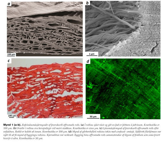
Figure 1. (a-b) SEM image of fish skin cross-section. (a) The fish skin is porous within but dense at the outer edges. Scale bar is 100 µm. (b) Fibers can be seen within the fish skin with more magnification. Scale bar is one µm. (c) Light microscopy image of fish skin cross-section. Pores can be seen within the fish skin . Scale bar is 100 µm. (d) confocal microscopy image of the fish skin surface. The structure consists of layers of perpendicular fibers. Scale bar is 50 μm.
Figure 2. SDS-PAGE analysis. (a) Protein size ladder. (b) Collagen I. (c) Collagen II. (d) Electrophoresis of fish skin protein extract yielded a faint band of roughly 115-130 kDa. (b-d) Themost noticeable band in the fish skin extract (115-130 kDa) overlaps with collagen I and partially with collagen II (n=3).
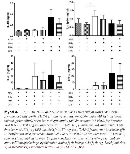
Figure 3. IL-6, IL-10, IL-12 and TNF-α were measured in the supernatant of moncytes or macrophages with an ELISA test. THP-1 cells were left untreated (48 hours, negative control, gray bars), cultured with the fish skin without treatment (48 hours), pre-treated with IFNγ (3 hours) and then stimulated with LPS (48 hours, positive control, white bars) with or without fish skin. THP-1 cells were also differentiated into macrophages with PMA pre-treatment (48 hours) before stimulation with LPS (48 hours, black bars) with or without fish skin. No significant difference was found within each condition with or without fish skin. Error bars represent standard error of the mean of 6 different experiments (n=6). *: P>0,05.
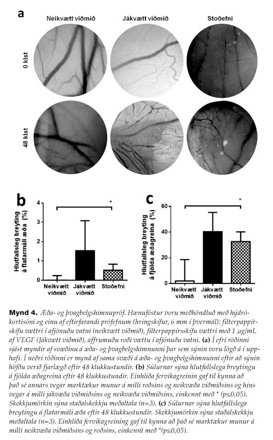
Figure 4. Chick CAM assay. Chick embryos received hydrocortisone and one of the following test materials (punches, 6 mm in diameter): hydrated filterpaper (negative control), hydrated filterpaper also with VEGF (1 µg/mL) (positive control), hydrated fish skin. (a) The top row shows images of the CAM where samples were placed in the beginning and the lower row shows the same area 48 hours later. (b) The columns represent the change in number of branching points after 48 hours. One-way ANOVA yielded a statistically significant difference between groups and the untreated control marked by * (p ≤ 0.01). Error bars represent standard error of deviation. (c) The columns represent the change in vascular area after 48 hours. One-way ANOVA yielded a statistically significant difference between the group that received fish skin and the untreated control marked by ** (p≤0.05). Error bars represent the standard error of deviation.