
10. tbl. 101. árg. 2015
Case of the month. An elderly woman with dyspnea and stridor
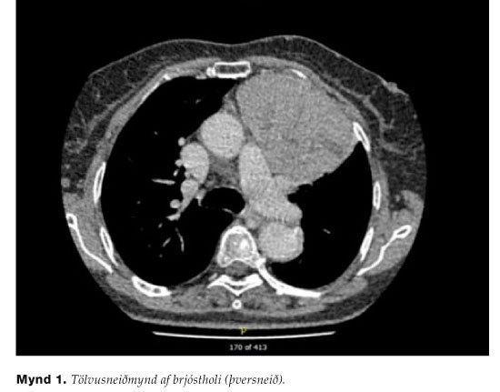
Figure 1. Computed tomography scan of the chest.
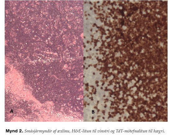
Figure 2. Microscopic picture of the tumor. H&E stain to the left and TdT immunoperoxidase staining to the right.
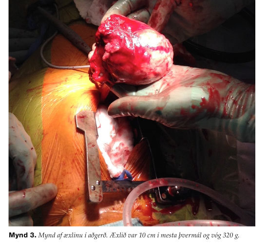
Figure 3. A photograph from the operation when the tumor was removed radically via a partial upper sternotomy.