
09. tbl. 102. árg. 2016
Cerebral arteriovenous malformations – overview
Ólafur Árni Sveinsson1, Ingvar H. Ólafsson2, Einar Már Valdimarsson3
1Department of Neurology Karolinska Hospital, Stockholm, Sweden, 2Department of Neurosurgery, University Hospital of Iceland, 3Department of Neurology, University Hospital of Iceland, Reykjavík, Iceland.
Cerebral arteriovenous malformations (AVMs) are uncommon but can cause intracerebral hemorrhage with grave disability or death. AVMs can even cause focal neurological symptoms, seizures and headache. The treatment of AVMs is complex. The most common treatment forms are microsurgery, stereotactic radiotherapy and endovascular embolization. The best treatment in each case can include a combination of the mentioned treatment forms. New studies indicate that no intervention is the best option in unruptured AVMs. In this article we discuss the epidemiology, diagnosis and treatment of cerebral AVMs.
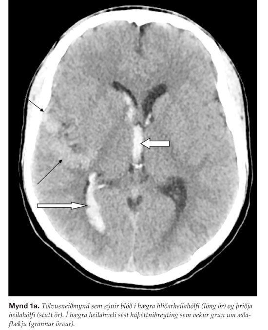
Figure 1a). CT without contrast shows intraventricular haemorrhage in the right lateral ventricle (long arrow) and third ventricle (short arrow). In the right hemisphere one can see a high density lesion which raised the suspicion of an AVM (narrow arrows).
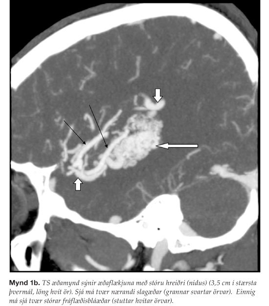
Figure 1b). CT angiography shows the AVM, with a large nidus (3.5 cm, in diameter, long narrow white arrow). Two feeding arteries (narrow black arrows) can be seen and furthermore two draining veins (short white arrows).
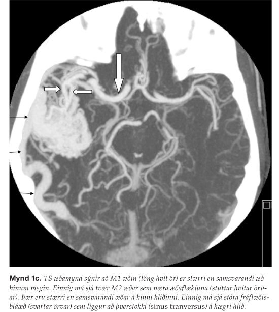
Figure 1c). CT angiography shows how the M1 artery (long white arrow) is larger than corresponding artery on the other side. Two feeding arteries (two short white arrows) can be clearly visualized. They are larger than corresponding opposite arteries. Additionally, one can depict a large vein that drains the AVM (narrow black arrows) which then drains in to the right transverse sinus.
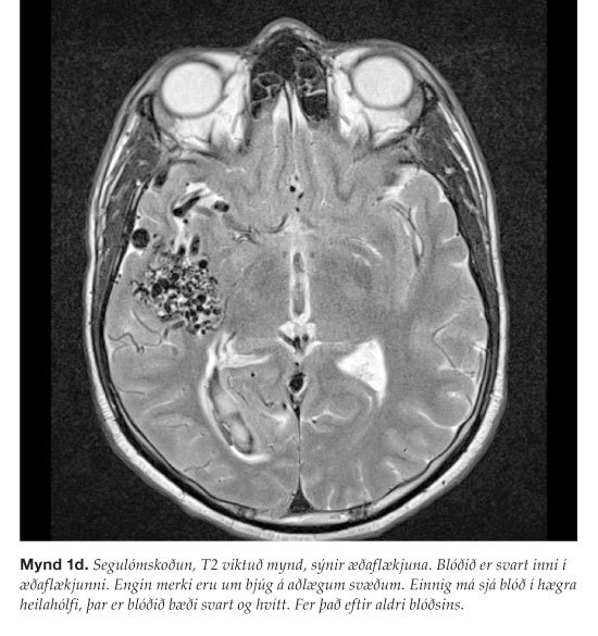
Figure 1d). T2 weighted MRI which clearly reveals the AVM. Blood is black inside the AVM. One can also see blood in the right ventricle. The blood is both black and white, depending on age. There are no signs of edema around the AVM.