
11. tbl. 99.árg. 2013
A female in her forties with dysphagia and chest pain. Esophageal leiomyoma
Tilfelli mánaðarins. Kona á fertugsaldri með kyngingarörðugleika og brjóstverki
A female in her forties with dysphagia and chest pain. Esophageal leiomyoma
Figures:
Fig. 1a. Computed tomography of the tumor (arrow).
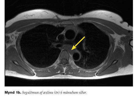
Fig. 1b. Magnetic resonance imaging og the tumor (arrow) six months later.
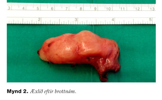
Fig. 2. Photography of the resected tumor
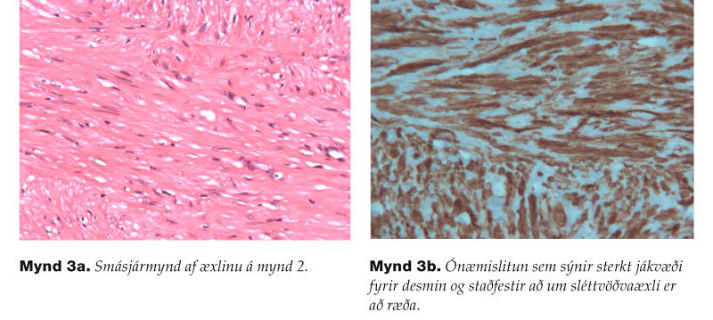
Fig. 3a. Microscopy of the tumor in Fig. 2 (H/E stain).
Fig. 3b. Tumor cells express desmin on immunostain that confirms the diagnosis of leiomyoma
Arnadottir H1, Gudjonsson H2, Sigurdardottir M3, Blöndal S4, Gudbjartsson T4
1Helsingborgs Lasarett, 2Gastrological department, 3Pathological division, 4Department of Cardiothoracic Surgery Landspítali, The University Hospital in Reykjavík
Key words: Leiomyoma, eosophagus, benign tumor, dysphagia, cheast pain.