
10. tbl. 102. árg. 2016
Case report: Intraocular foreign body
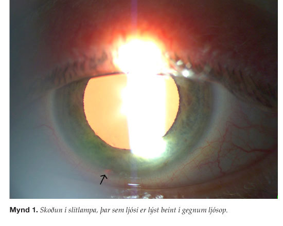
Figure 1. Slit lamp examination of the left eye, transillumination.
Figure 2. Fundoscopy of the left´s eye.
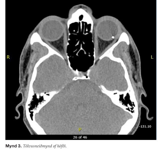
Figure 3. Computed tomography (CT) of orbits.
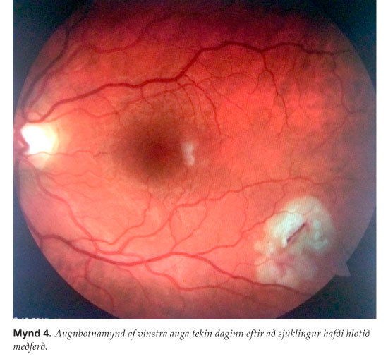
Figure 4. Fundus photography of the left eye the day after pars plana viterectomy.
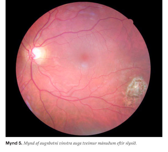
Figure 5. Fundus photography of the left eye two months after the accident.
Elín Björk Tryggvadóttir1, Óskar Jónsson1,2, Gunnar Már Zoega1,2
1Department of Ophthalmology, Landspitali University Hospital, 2Sjónlag Eye Center.