
04. tbl. 99. árg. 2013
Cardiac perforation following pacemaker implantation – a case series from Iceland
Introduction: Perforation of the heart is a serious complication following pacemaker implantation that can cause life threatening bleeding and cardiac tamponade. Here we describe five cases that were diagnosed in Iceland during a four year period.
Materials and methods: This population-based case series includes five patients diagnosed with cardiac perforation following pacemaker insertion at Landspítali and Akureyri Hospital from January 1, 2007 to December 31, 2010. The mode of detection, treatment given and outcome were studied.
Results:Altogether five patients (mean age 71 years, three females) were diagnosed with cardiac perforation in Iceland during the study period, one in 2008 and four in 2009. Chest pain was the most common presenting symptom (n=4) and no patient had acute cardiac tamponade. In all five cases the diagnosis was obtained with computed tomography scan or echocardiography. No perforation was detected intraoperatively but four of the cases were diagnosed within three weeks of the operation. Three patients were treated with surgical evacuation of blood via sternotomy and suture of the perforation. In the other two cases the pacemaker leads were removed in the operating room with trans-oesophageal echocardiographic guidance. Four patients survived the treatment and were discharged but one died of pneumonia in the intensive care unit.
Conclusion: Cardiac perforation is a serious complication and should be kept in mind in patients with chest pain following pacemaker insertion.
Sverrisson ITh1, Hognason J2, Vidarsdottir H1, Gottskalksson G2, Gunnarsson GTh3, Sverrisson JTh3, Gudbjartsson T1,4
1Departments of Cardiothoracic Surgery and 2Cardiology National University Hospital of Iceland, Reykjavík. 3Department of Internal Medicine, Akureyri Hospital, 4Faculty of Medicine, University of Iceland.
Figure 1. A CT scan (patient no. 3) that shows how the pacemaker wire has perforated the right ventricle (arrow).
Figure 2. A picture taken intra-operatively (patient no. 4). The pacemakerwire sticks out through the anterior wall of the right ventricle (arrow).
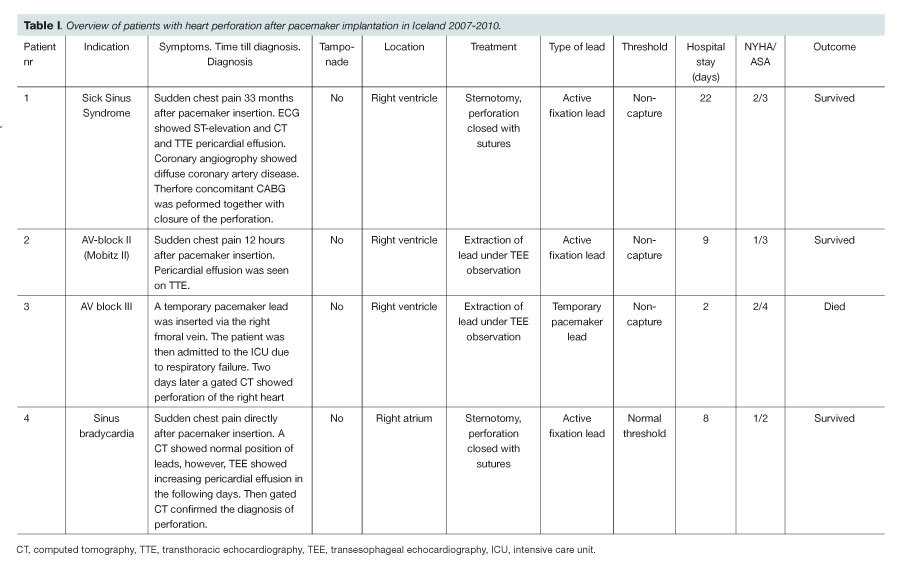
Table I. Overview of patients with heart perforation after pacemaker implantation in Iceland 2007-2010.
See table I here.