
11. tbl. 100. árg. 2014
Pulmonary alveolar proteinosis - a case report
Próteinútfellingar í lungnablöðrum meðhöndlaðar með lungnaskoli
Pulmonary alveolar proteinosis (PAP) is a rare lung disease of unknown origin, where an amorphous lipoprotein material accumulates in the alveoli of the lungs. We describe a young male with a four month history of progressive dyspnea, low grade fever, hypoxemia and weight loss. Chest X-ray showed diffuse interstitial and alveolar infiltrates in both lungs. The diagnosis of PAP was confirmed with trans-bronchial lung biopsy. Because of a deteriorating clinical course a whole lung lavage was performed. Under general anesthesia, both lungs were lavaged with warm saline in two different sessions with good results. Two years later the patient is almost free of symptoms and lung function has markedly improved.
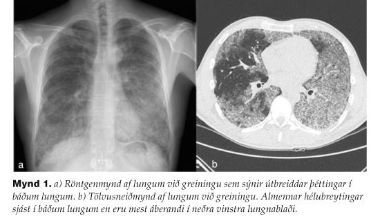
Figure 1. a) Chest X-ray at diagnosis showing diffuse infiltrations in both lungs. b) CT scan of the thorax at diagnosis showing ground glass consolidations that are most prominent in the left lower lobe.
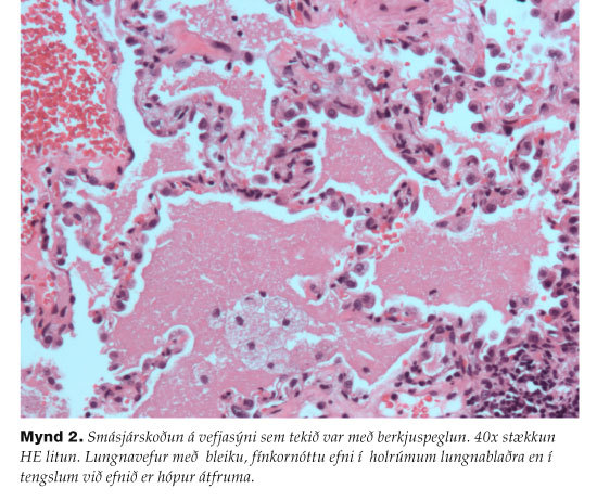
Figure 2. Microscopy of a lung biopsy taken with bronchoscopy(40x magnification HE stain). Alveolar spaces are filled with eosinophilic proteinaceous material. Foamy macrophages are present within the material.
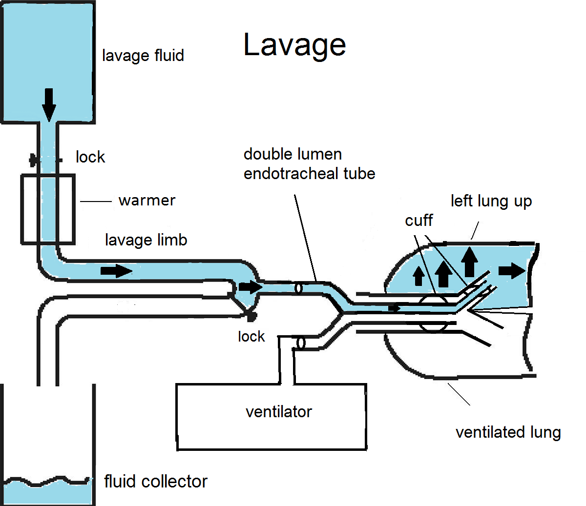
Figure 3a, Figure 3b. Whole lung lavage setup and equipment. The lavage fluid is placed on an IV pole and the fluid runs through a warmer. A lock is put between the fluid and the warmer to control volume instilled to the lung. Only one lung is ventilated while the other is being lavaged. There is a lock on the drainage limb to control the timing of drainage into the fluid collector. Figure: Felix Valsson, adopted and changed from a figure in reference 5.
Figure 4. Samples of bronchial lavage fluid from the left lung, showing gradually more dilute fluid as more saline was introduced into the lung.
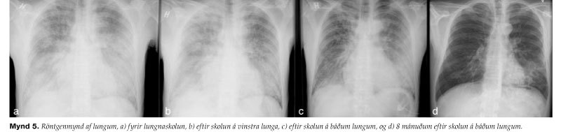
Figure 5. Chest X-ray a) before WLL, b) after WLL on the left lung, c) after WLL on both lungs, and d) 8 months after WLL.